Key types of microscopes and their features
2024-01-19
Microscopes are optical instruments that magnify small objects or details, allowing for the observation of structures that are not visible to the naked eye. Microscopes have played a crucial role in scientific research, medical diagnosis, and various fields where the examination of tiny details is necessary. There are different types of microscopes, each designed for specific applications. Here are some key types of microscopes and their features:
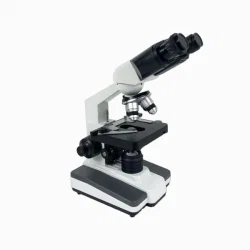
1. Optical Microscopes:
- Light Source: Optical microscopes use visible light as the source of illumination.
- Magnification: They offer magnification through a combination of objective lenses and eyepieces.
- Common Types:
- Compound Microscope: Uses multiple lenses to provide high magnification for observing thin sections of specimens, often used in biology and histology.
- Stereo Microscope (Dissecting Microscope): Provides a three-dimensional view of larger specimens at lower magnification, commonly used for dissection and inspection.
2. Electron Microscopes:
- Source of Illumination: Electron microscopes use a beam of electrons instead of visible light.
- Magnification: They offer extremely high magnification, enabling the observation of ultra-fine details.
- Common Types:
- Transmission Electron Microscope (TEM): Transmits electrons through thin specimens to create detailed images of internal structures.
- Scanning Electron Microscope (SEM): Scans a focused electron beam across the surface of a specimen to create 3D images.
3. Scanning Probe Microscopes:
- Probe Mechanism: These microscopes use a physical probe to scan the surface of a specimen.
- Magnification: They provide high-resolution images at the atomic and molecular levels.
- Common Types:
- Atomic Force Microscope (AFM): Measures the interaction forces between a sharp probe and a specimen's surface to create images.
4. Fluorescence Microscopes:
- Fluorescent Labels: Fluorescence microscopes use fluorescent dyes or labels to enhance contrast and observe specific structures or molecules.
- Applications: Commonly used in biology and medicine for studying live cells and cellular components.
5. Confocal Microscopes:
- Optical Sectioning: Confocal microscopes use a pinhole to eliminate out-of-focus light, allowing for optical sectioning and three-dimensional imaging.
- Applications: Useful for studying thicker specimens and creating detailed 3D reconstructions.
6. Digital Microscopes:
- Digital Imaging: These microscopes incorporate digital cameras for imaging, enabling the capture and storage of digital images and videos.
- Applications: Suitable for educational purposes, routine inspections, and documentation.
7. Inverted Microscopes:
- Design: Inverted microscopes have the light source and objectives positioned below the specimen stage, making them suitable for observing cells or tissues in culture dishes.
- Applications: Widely used in cell biology, microbiology, and tissue culture studies.
8. Phase Contrast Microscopes:
- Contrast Enhancement: Phase contrast microscopes enhance the contrast of transparent and colorless specimens by exploiting differences in refractive index.
- Applications: Useful for observing live cells and unstained biological specimens.
9. Polarizing Microscopes:
- Polarized Light: Polarizing microscopes use polarized light to study the optical properties of materials, such as minerals and crystals.
- Applications: Commonly used in geology, mineralogy, and materials science.
10. Darkfield Microscopes:
- Contrast Enhancement: Darkfield microscopes use oblique illumination to enhance contrast by illuminating the specimen against a dark background.
- Applications: Useful for observing transparent or low-contrast specimens.
Microscopes are essential tools in various scientific and medical disciplines, enabling researchers and professionals to explore the microscopic world with precision and detail. The choice of microscope depends on the specific requirements of the application, including the type of specimen, desired magnification, and imaging techniques.